 ObjectExplorer
Lab and life science
Genomics fasta fastq bed gff3 gtf vcf bcf sam bam cram, lab files gb gbkembl fcs ab1 dna bigwig bigbed, microscopy ome.tif czi nd2 lif, mass spectra mzml, and molecules pdb ent cif mmcif mol sdf mol2 xyz
Sequences, traces, cell counts, microscope stacks and molecules open as what they hold, read on your machine: no Python, no plug-in. Which reader a file needs, its first bytes decide.
Genomics
Every one of these opens as a table, a row per record, its text a click away:
| File | A row for each, with |
|---|---|
FASTA fasta fa fna faa | sequence: identifier, description, length, sequence |
FASTQ fastq fq | read, with its quality and the mean quality |
BED bed | region, in UCSC's column names, BED3 to BED12 |
GFF3 and GTF gff3 gff gtf | feature: the nine columns, and a column for each attribute |
VCF vcf, and BCF, its binary bcf | variant: the #CHROM columns and one per sample |
SAM sam, BAM bam, CRAM cram | alignment: the eleven fields and a column per optional tag |
VCF is the Variant Call Format, SAM the Sequence Alignment Map (BAM is its binary, CRAM the same compressed against the reference), GFF the General Feature Format, GTF the Gene Transfer Format and BED the Browser Extensible Data of UCSC's (the University of California, Santa Cruz's) genome browser. A record that is broken still shows as a row, with a last column saying what is wrong. A BAM's or a CRAM's structure mode lays out its header, references and compressed blocks. A .vcf that is a contact card opens as one — see records and trees.
CRAM 3.0 is read. A CRAM compressed with a method of CRAM 3.1 says which; a read whose bases are kept only as differences from a reference sequence the file does not hold shows * for them, and its problem column names that reference.
Lab files
GenBank and EMBL (the European Molecular Biology Laboratory) entries, flow cytometry files (FCS, the Flow Cytometry Standard), Sanger traces in ABIF (Applied Biosystems' Interchange Format), SnapGene's plasmid maps, and the bigWig and bigBed tracks of UCSC's genome browser are files of several tables:
| File | Its tables |
|---|---|
GenBank gb gbk genbank gbff gpff gp | records, features |
EMBL embl | records, features |
FCS 2.0 to 3.2 fcs lmd | events, parameters, keywords |
Sanger trace ab1 abi abif fsa | bases, traces, tags |
SnapGene dna | sequence, features, primers |
bigWig and bigBed bigwig bb bigbed | intervals or entries, a zoom level each, chromosomes |
A file of these opens as the list of its tables, as an SQLite file does (a Sanger trace drawn first, below), and its notebook queries one by its name after the file's path: SELECT * FROM 'runs/sample.fcs/events'.
A GenBank or EMBL feature carries its record, its type, its location as written and its first and last base. A flow cytometry file's events are a column a parameter, by its name (FSC, SSC, FL1 …), as the instrument stored them.
A Sanger trace opens drawn in Signals: its four traces a lane each, the bases called at their places, counted in scans rather than seconds — the first 500 scans, then more with the window's lengths and the slider, as a recording is moved.
A bigWig named .bw, the name of an SGI (Silicon Graphics) picture, is told by its bytes and opens as the track it is.
Microscopy
| File | From |
|---|---|
OME-TIFF ome.tif ome.tiff | the Open Microscopy Environment's tools |
CZI (Carl Zeiss Image) czi | Zeiss |
ND2 nd2 | Nikon |
LIF (Leica Image File) lif | Leica |
A microscope's stack opens slice by slice, the way a volume does, with a card of its facts beside it: its size, its axes, its channels, how far apart its pixels are. The row over the slider shows All the channels together, each in its own colour, or one alone over its own range. A slider for each further axis is named after it — slice, time, position, wavelength — and Axial, Coronal and Sagittal appear when the stack has depth. The Table holds its planes, its header and the metadata the microscope wrote.
An OME-TIFF is told from a plain TIFF by the description it carries. A CZI's planes compressed with JPEG, LZW or JPEG XR are named, not drawn; an ND2 of Nikon's oldest kind, which is a JPEG 2000 file, opens as that picture.
Mass spectra
An mzML file, a mass spectrometer's run, opens as its table: a row a spectrum, with its id, its level, its scan time, its polarity, its kind and its base peak. A file older than mzML 1.0 is refused, and says so.
Molecules
| File | What is drawn |
|---|---|
Protein Data Bank (PDB) pdb ent brk, mmCIF cif mmcif mcif | its first model |
a crystal's CIF (Crystallographic Information File) cif | its unit cell, filled out by its symmetry |
MDL's molfile mol, and a structure-data file (SDF) of them sdf sd | every record side by side, up to 400 |
Tripos MOL2 mol2 ml2 | every molecule side by side, up to 400 |
XYZ and extended XYZ xyz extxyz | its first frame |
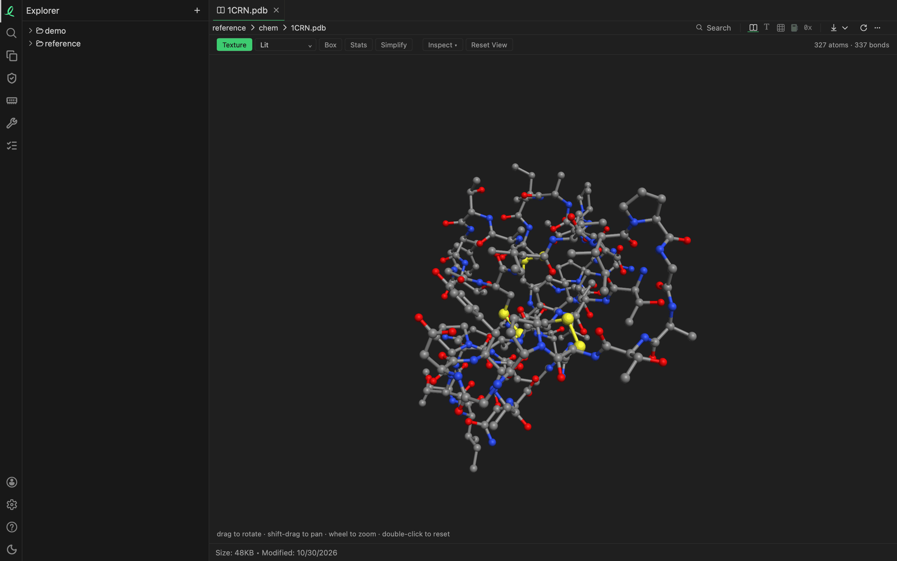
A molecule opens drawn ball and stick, in 3D: an element a colour, a double or a triple bond two or three thinner sticks, and the counts of its atoms, bonds and models over it. Drag to turn it, Shift-drag to move it, the wheel to zoom, a double click to reset — the same view as a 3D model. Bonds the file does not list are found from how close the atoms stand.
Its text is next, then its atoms as a table, a row an atom; an SDF's table is a row a record, with its name, atoms, bonds, formula and weight, and a column for each data item it carries. A .pdb that is a Palm ebook opens as an ebook; the bytes tell them apart.
Next: medical and health.